
Presentation
Chronic pain in the back of the right ankle with no history of trauma.
Patient Data
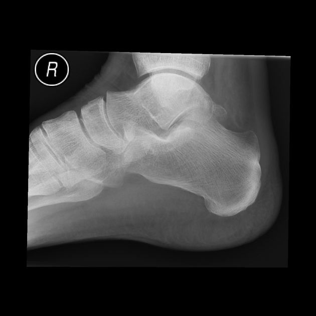
There is soft tissue density noted in the region of Kager's fat pad/ retrocalcaneal soft tissue suggestive of retrocalcaneal bursitis.There is also minimal thickening of the Achilles tendon suggestive of Achilles tendinopathy.
The bony structures of the ankle joint appear normal. The articular surfaces are intact. Alignment of the ankle mortice is anatomical. No joint effusion is evident. There is soft tissue swelling overlying the distal Achilles tendon.
Further examination of the soft tissues by ultrasound has been performed.


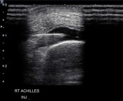
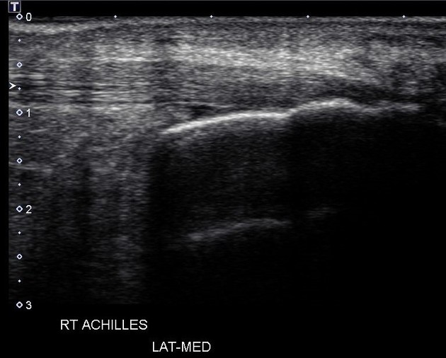
The Achilles tendon shows thickening proximal to the insertion. There is moderate inflammation of the bursa at the calcaneal insertion. There is no calcification at the calcaneal insertion. There is no evidence of a discrete tear or focal injury. The remaining subcutaneous soft tissue echogenicity and morphology is unremarkable.
Under sterile conditions 1.0 ml Celestone 5.7 mg/ml (Chronodose) and 1.0 ml Marcaine 0.5% was injected into the retrocalcaneal bursa. .
Case Discussion
There are appearances in keeping with retrocalcaneal bursitis and mild Achilles tendinopathy.


 Unable to process the form. Check for errors and try again.