
Presentation
Incidental left temporal lobe lesion found on outside imaging.
Patient Data
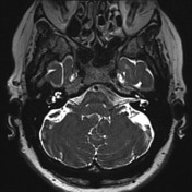


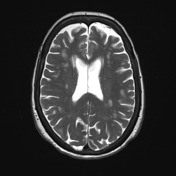
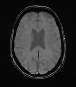
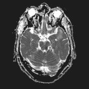
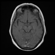
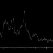
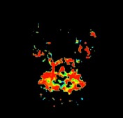
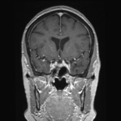

In the subcortical white matter of the left temporal pole are a number of cystic spaces surrounded by high T2 signal which does not involve the overlying cortex. The cysts are intimately associated with MCA branches, and the largest cyst appears to have a thin communication through the cortex to the adjacent vessel. MRS and CBV are normal and no enhancement is present.
Case Discussion
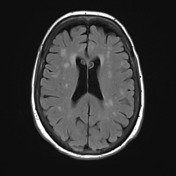
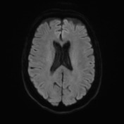
This patient has now been followed for 5 years. The cysts remain the same, and at most there has been minor reduction in surrounding T2 signal.
Although without histology it is difficult to prove the diagnosis, the location, the stability over time and particularly the relationship of the largest cyst to the MCA branch overlying it are very suggestive of the diagnosis of anterior temporal lobe perivascular cysts.


 Unable to process the form. Check for errors and try again.