
Presentation
Headache, new hypertension, vertigo and nystagmus.
Patient Data


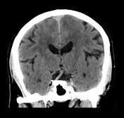
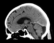
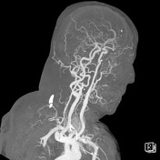

No acute intracranial pathology identified on non-contrast CT. Hypodense foci in the left cerebellar hemisphere in keeping with prior infarctions. No vascular abnormality is identified in the neck vessels and circle of Willis.
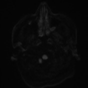
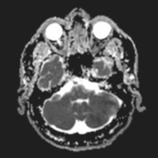
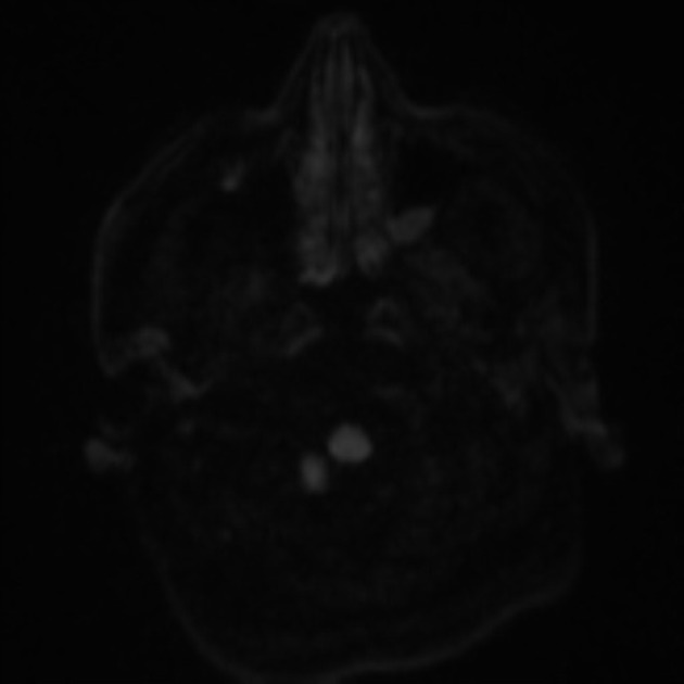
There are numerous acute lacunes in the deep white matter of the posterior left cerebral hemispheres, and true on the left cerebellar hemisphere. Grossly preserved flow voids in the basilar artery. Findings are consistent with an embolic stroke.

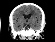
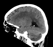
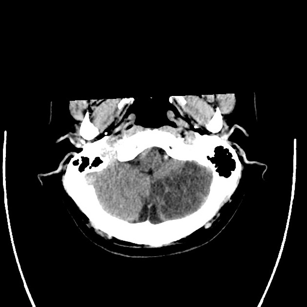
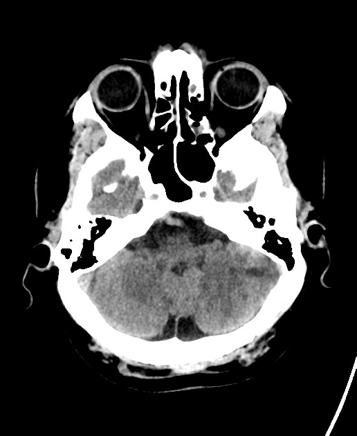
Well-defined hypoattenuation involving the most inferior aspect of the left cerebellar hemisphere is in keeping with the left PICA vascular territory infarct. There is no significant mass effect over the brainstem and fourth ventricle. No hemorrhage transformation. Supratentorial ventricles and basal cisterns are unremarkable. White matter hypoattenuation areas are nonspecific, most likely representing chronic small vessel ischemia, in a number distribution over the expected for the patient's age group. No evidence of new infarcts. The lacunes seen on the MRI are not well appreciated on CT.
Case Discussion
Features are consistent with multiple small embolic cerebral infarcts involving the left MCA, left PCA and left PICA territory. The last, further characterized by a more prominent territorial infarct.
Note also that on the CTA there is a fenestrated basilar artery, which is an anatomical variant.


 Unable to process the form. Check for errors and try again.