
Presentation
Slow growing masses on the neck with multiple resections done over 4 decades.
Patient Data
Age: 65 years
Gender: Male
From the case:
Plexiform neurofibroma


Download
Info

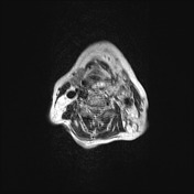
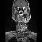
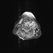
There are multiple confluent enhancing lesions predominantly on the left side of the neck and in the upper mediastinum which are responsible for an asymetrical stenosis of the pharynx and trachea. Notice the target sign on T2 sequence typical for neurofibromas.
Case Discussion
This patient underwent several surgical resections throughout last decades (and also recent chemotherapy treatment) with histologically confirmed diagnosis of plexiform neurofibroma (see article Neurofibromatosis type I). There were also signs of malignant transformation documented.


 Unable to process the form. Check for errors and try again.