
Presentation
Progressive swelling of the right eye lid since 11 years.
Patient Data
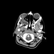

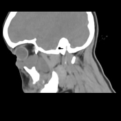
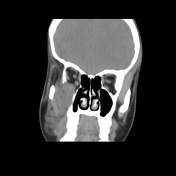
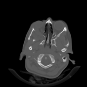
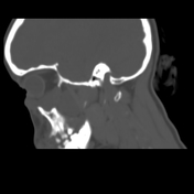
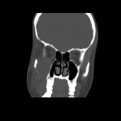

An extraconal soft tissue mass is present along the inferior aspect of the right orbit, centered in the infraorbital canal and extending into the inferior orbita fissure and infratemporal fossa.


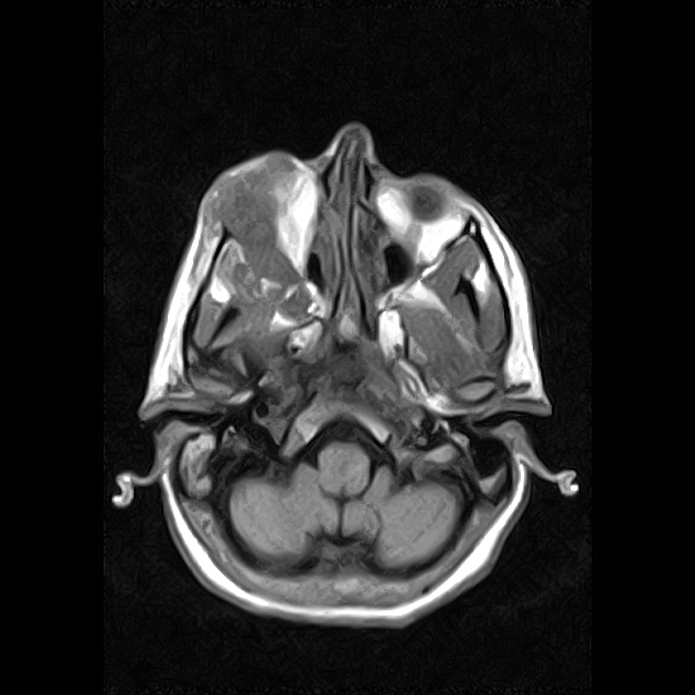
A lobulated and serpentine extra-conal soft tissue mass lesion is seen creeping along the inferolateral aspect of the right orbit and extending to the right infratemporal fossa through a widened inferior orbital fissure with associated widening of the foramen rotundum and pterygomaxillary fissure with possible extension backward into the pterygopalatine ganglion and its branches within the pterygopalatine fossa. The lesion measures about 5.4 X 2.5 x 4 cm in its maximal axial and craniocaudal dimensions respectively. It shows heterogeneous intermediate signal in T1 turning into bright signal in both T2 and T2 STIR images with mild homogeneous post-contrast enhancement. The intra-orbital component is seen encroaching upon the intraorbital fat closely related to the inferior aspect of the eye globe with no right orbital proptosis as well as indenting the inferior and lateral recti muscles with no significant displacement. The lesion extends anteriorly and inferiorly within the right lower eyelid. The infra-temporal component is seen related to the mandibular ramus and lateral pterygoid muscles and shows subcutaneous extension seen superficial to the right masseter as well as the temporalis muscles.
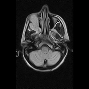
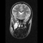
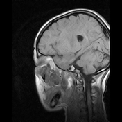
The limited axial T2 and FLAIR study of the brain shows a small focus of bright signal within the left basal ganglia.
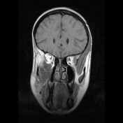
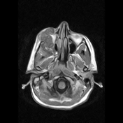
Normal symmetrical spherical configuration of both eye globes. No left sided intra- or extra-conal masses could be detected. Preserved signal with comparable appearance of the left extra-ocular muscles bilaterally with normal course and caliber. Normal MR caliber, course, and signal of both optic nerves. No masses seen. Normal MR appearance of the superior ophthalmic veins. No sellar, supra or para-sellar masses. Preserved MR signal of both cavernous sinuses.
Case Discussion
A lobulated and serpentine extra-conal soft tissue mass lesion creeping along the infero-lateral aspect of the right orbit and extending to the right infra-temporal fossa through a widened inferior orbital fissure; most likely representing a plexiform neurofibroma of the infra-orbital nerve with left basal ganglionic small demyelinating lesion is likely unidentified bright object (UBO) related to neurofibromatosis type I (NFI); for clinical correlation and chromosomal analysis / genetic counseling.
Misdiagnosis of the facial plexiform neurofibroma with venolymphatic malformation is not uncommon.


 Unable to process the form. Check for errors and try again.