
Presentation
Recent subarachnoid hemorrhage
Patient Data
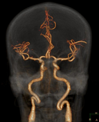
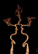
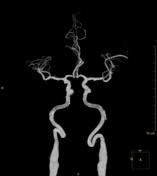
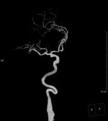

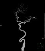
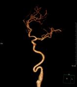
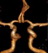

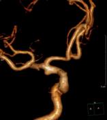

Broad based bulge (blood blister like) projects at the dorsal/lateral wall of the supraclinoid portion of the right internal carotid artery just below to its bifurcation. It measures about 4mm in maximum diameter. It has a wide neck.
Case Discussion
Blood blister-like aneurysm is a broad-based bulge at a nonbranch point of a vessel.
Clinical presentation
Middle-aged patients, angiographically negative for subarachnoid hemorrhage.
Radiographic features
shape: blood blister-like or half-domed shallow outpouching with wide neck
common sites: supraclinoid ICA (dorsal wall), less likely MCA, ACA, basilar artery
size: usually small (<6 mm) mean (3 mm)
rapid change in size and morphology on follow up angiograms
CT angiography: often negative
digital subtraction angiography: the best diagnostic tool
Differential diagnosis
vessel infundibulum
vasospasm
atherosclerotic vascular disease


 Unable to process the form. Check for errors and try again.