
Presentation
Seizures. History of old cranial gunshot injury.
Patient Data


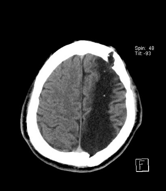
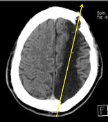
CT soft tissue window. The hypodense track of encephalomalacia delineates the bullet track with a large intracranial left frontal bone fragment.
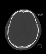
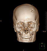
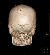
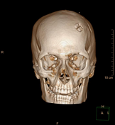
Bone window demonstrates internal beveling at the entry site at the left posterior parietal calvarial bone with the exit site seen at the left frontal calvarial bone.
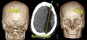

First image: bevelled bullet entry at the left posterior parietal bone.
Second image: larger bullet exit at the left frontal bone.
Third image: bullet track.
Case Discussion
The patient suffers from the remote sequelae of a former cranial gunshot injury.
Cranial gunshot injuries are a form of penetrating traumatic brain injury, which are much less common than blunt traumatic brain injuries.


 Unable to process the form. Check for errors and try again.