
Presentation
Presented with recurrent UTI and renal calculi. Found to have hypercalcemia.
Patient Data

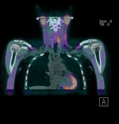
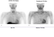
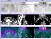
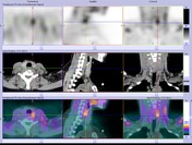
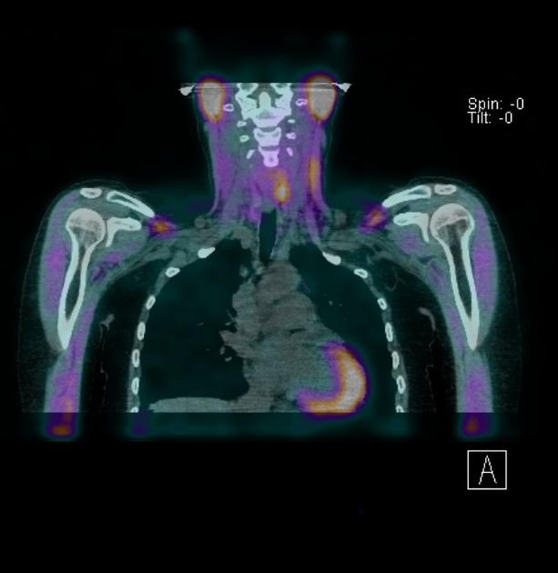
Static and SPECT images were acquired at 15 min and 3 hours. Delayed CT- SPECT was performed.
Initial images show mild focal uptake in the region of the thyroid left upper and right lower poles. No other abnormal extra-thyroid uptake is detected in the rest of the neck or mediastinum. Delayed images show locally retained uptake corresponding to a soft tissue nodule posterior to the left upper pole and another little nodule posterior to the right lower pole. Mild uptake in sub centimeter left supraclavicular lymph nodes is non-specific. Mild uptake in the left anterior mediastinum is probably within physiological limits.
Case Discussion
Scintigraphic findings are consistent with two parathyroid adenomas posterior to the left thyroid upper pole and right lower pole. These were confirmed histologically after resection:
MICROSCOPIC DESCRIPTION: 2. The section shows parathyroid tissue in which there is a small irregularly shaped nodule. This is composed predominantly of chief cells with smaller components of oxyphil and water clear cells noted. The nodule is enclosed within a thin fibrous capsule. No evidence of capsular or vascular invasion is seen. A small amount of normal appearing parathyroid tissue is present at one edge external to the capsule. The features are consistent with parathyroid adenoma. no evidence of malignancy is seen. 3. The section shows parathyroid tissue in which there is a small nodule enclosed within a thin fibrous capsule. This is composed almost entirely of chief cells. A small amount of normal parathyroid tissue is present at one edge external to the capsule. The features are consistent with parathyroid adenoma. No evidence of malignancy is seen. 6. The section shows a single small unremarkable parathyroid. No evidence of tumor is seen.
DIAGNOSIS: Features consistent with parathyroid adenoma.


 Unable to process the form. Check for errors and try again.