
Presentation
Follow-up of a known condition.
Patient Data


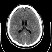
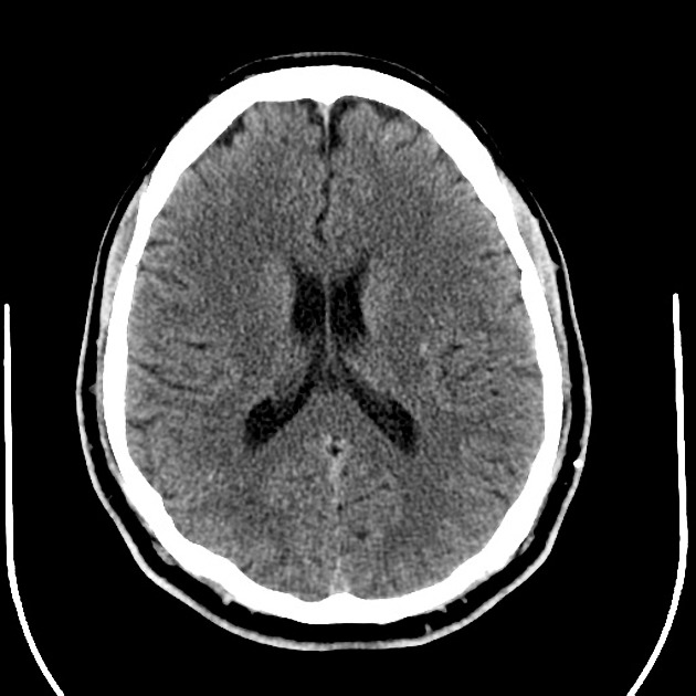
There are multiple subtle ill-defined hyperdense foci scattered through the brain, some of which show faint enhancement. The largest discrete lesion is probably in the right middle cerebellar peduncle, measuring approximately 7 mm in size. No acute hemorrhage (away from the hyperdense lesions) or mass effect is detected. The ventricles and sulci are normal in size.
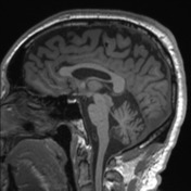
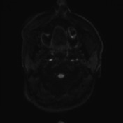
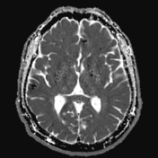

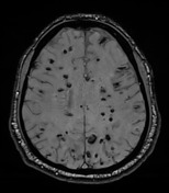

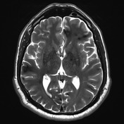
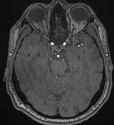
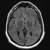
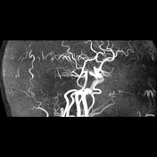
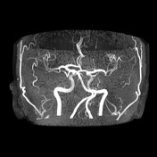
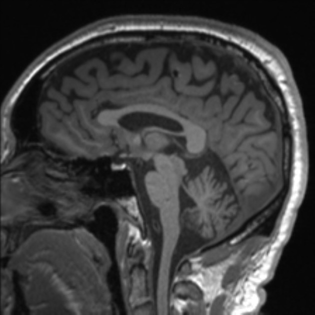
Multiple nodular foci of low signal and marked blooming artefact are again demonstrated scattered through the brain, brainstem, and cerebellum, with at least two new foci when compared to the last MRI brain. Bilateral subcortical low signal consistent with hemosiderin staining are again demonstrated, more significant in the right frontoparietal region. No evidence of acute hemorrhage. Ventricles and basal cisterns are normal in appearance. MRA demonstrates no malformation, aneurysm, stenosis or other vascular abnormality.
Case Discussion
This case demonstrates an exuberant presentation of multiple cerebral cavernous malformations.
Although no further clinical details were available, this case raises the possibility of familial multiple cavernous malformation syndrome.
This condition is inherited in an autosomal dominant manner, although cases caused by de novo gene mutations are recognized.


 Unable to process the form. Check for errors and try again.