
Presentation
Seizure
Patient Data


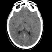
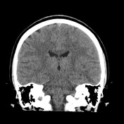
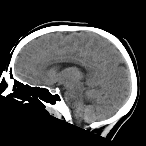
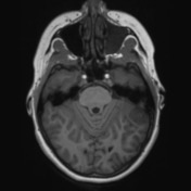
Low attenuation and mass effect is present in the posteroinferior aspect of the left temporal lobe.
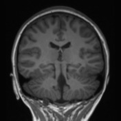
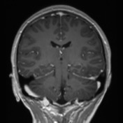
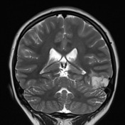
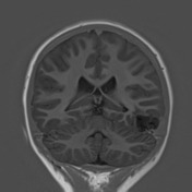
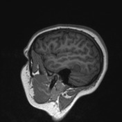
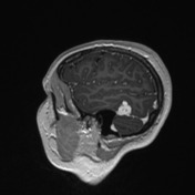
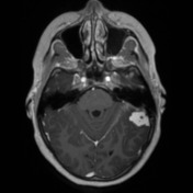
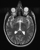

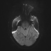
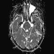
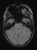

A high T2, low T1, vividly enhancing mass abutting the tentorium on the left and invaginating into the posterior temporal lobe is present. It contains prominent flow voids appearing to arise from, or drain to, the dura near the transverse sinus.
Case Discussion
The patient went on to have a resection.
Histology
MICROSCOPIC DESCRIPTION:
Paraffin sections show fragments of an intensely vascular, moderately hypercellular tumor with a meningeal attachment. Vascular spaces vary from large caliber thin-walled sinuses down to capillaries. The latter enclose lobules of cells with features consistent with stromal cells. No mitotic figures or areas of necrosis are identified.
Immunohistochemistry shows negative staining for EMA, inhibin, CD34, GFAP, CAM5.2, S-100, tyrosinase and ALK-1.
DIAGNOSIS: Hemangioblastoma (WHO grade I).
Discussion
This case exemplifies the difficulty in reconciling various factors (location, appearance, demographic) when appearances are unusual. It also highlights the fact that their pial origin makes a distinction between intra- vs extra-axial difficult.


 Unable to process the form. Check for errors and try again.