
Presentation
Headache and visual disturbance in the last days.
Patient Data


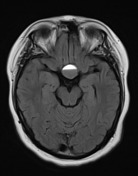
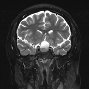
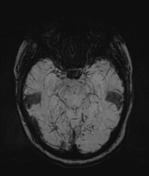
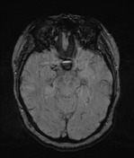
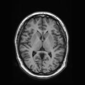
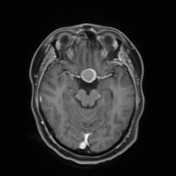
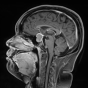
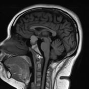

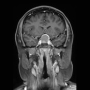
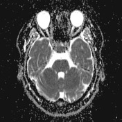
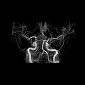
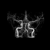
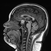
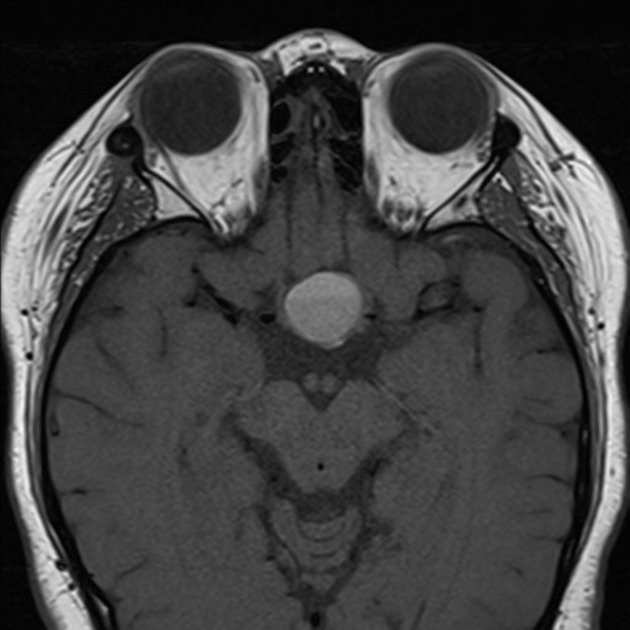
Enlarged sella with a pituitary adenoma with a suprasellar extension due to a hemorrhagic component. The tumor indents the cavernous sinus on the right without further invasion. The suprasellar component has an intrinsic T1 signal and susceptibility artefact consistent with hemorrhage, also with a hematocrit level within. This pushes the optic chiasm superiorly. Flow void of the ICA is preserved bilaterally. Brain is otherwise normal. Mucosal thickening of the left maxillary and right frontal paranasal sinuses.

MACROSCOPY:
- "Pituitary": A piece of soft tan tissue 3mm. DIAGNOSIS: Pituitary adenoma.
- "Pituitary lesion": Pale pink tissue fragments 5mm in aggregate.
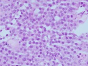
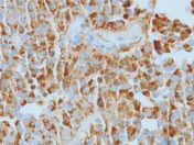
MICROSCOPY: 1&2. Sections show a hypercellular tumor composed of solid sheets, nests and trabeculae. Tumor cells contain scant granular eosinophilic cytoplasm, round nuclei with finely granular chromatin and inconspicuous nucleoli. No mitoses or necrosis are seen. No normal anterior pituitary tissue is included. No brain invasion is seen.
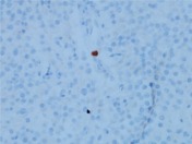
Immunohistochemical results show positive Prolactin and CAM5.2 staining, whilst LH, FSH, GH, ACTH and TSH pituitary hormones are negative. The Topoisomerase proliferation index is 1%.
CONCLUSION: Pituitary tumor: Densely granulated Prolactin secreting pituitary adenoma.
Case Discussion
Typical imaging features and clinical presentation of pituitary apoplexy in a further confirmed prolactinoma.


 Unable to process the form. Check for errors and try again.