
From the case:
Cholesterol granuloma


Download
Info

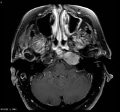
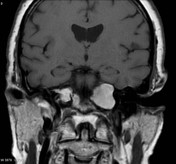
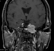
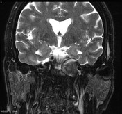
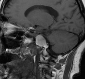
MRI through the base of skull demonstrates a well circumscribed mass at the left petrous apex. It shows striking high T1 signal and demonstrates no convincing enhancement or fat suppression on fat suppressed post contrast images.
T2 signal is heterogeneous with a thin rim of signal drop out peripherally.
Case Discussion
Appearances are consistent with a cholesterol granuloma also known in this location as a giant cholesterol cyst.


 Unable to process the form. Check for errors and try again.