
Presentation
Both side groin pain. Pain worsened on weight bearing.
Patient Data
Age: 40 years
Gender: Female
From the case:
Avascular necrosis of hip (MRI)
Download
Info
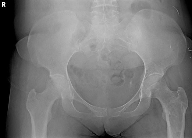
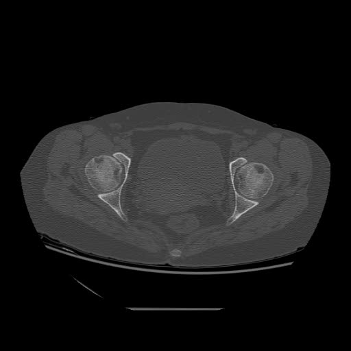
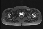
The bilateral joint spaces shows mild narrowing with femoral articular surface irregularity.
There is a well defined expansile lesion with sclerotic borders and narrow zone of transition noted in the left proximal femoral diaphysis. No periosteal reaction. No pathological fracture
From the case:
Avascular necrosis of hip (MRI)


Download
Info

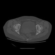
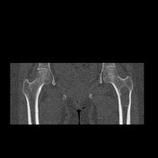
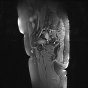
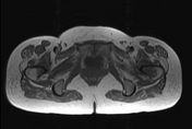
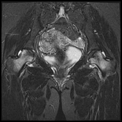
Bilateral femoral heads show early collapse with subchondral fracture and diffuse sclerosis.
The proximal femur shows diffuse edema.
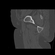
Left proximal femur simple bone cyst. No abnormal soft tissue component within.
Bilateral small volume hip joint effusion also seen.


 Unable to process the form. Check for errors and try again.