
Presentation
Presentation with initial left sided hemiplegia for 2 hours and quickly developing coma.
Patient Data
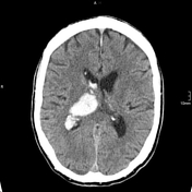


Hyperacute intracranial hemorrhage affecting the right thalamus with extension into the ventricle, hyperdense on CT.

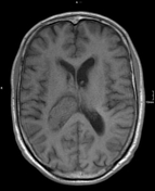
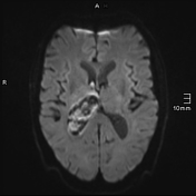
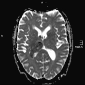
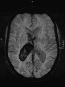
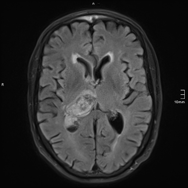
Hyperacute intracranial hemorrhage affecting the right thalamus with extension into the ventricle. Imaging at 3T MRI.
T1 isointense signal.
T2 FLAIR partially isointense (representing the clot), partially slightly hyperintense (representing a halo of serum outside of the clot).
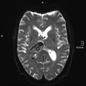
DWI/ADC demonstrating restricted diffusion of the clot, facilitated diffusion of the halo (serum adjacent to the clot).
SWI demonstrating signal loss.
Case Discussion
Hyperacute intracranial hemorrhage affecting the right thalamus with extension into the ventricle. In the elderly, the most common cause of this type of hemorrhage is poorly controlled hypertension. In younger patients, or those without hypertension, an underlying vascular or potentially neoplastic lesion should be sought.


 Unable to process the form. Check for errors and try again.