
Presentation
This patient is from a nursing home. Right sided weakness developed rapidly a few weeks ago. Non contrast CT demonstrated low density region.
Patient Data
Age: 70 years
Gender: Female
Download
Info

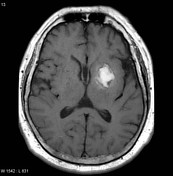
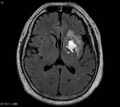
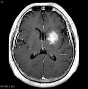
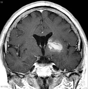
A region of irregular peripheral enhancement involves the left lentiform nucleus and internal capsule.
Download
Info

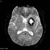
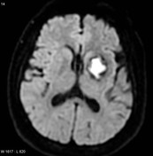
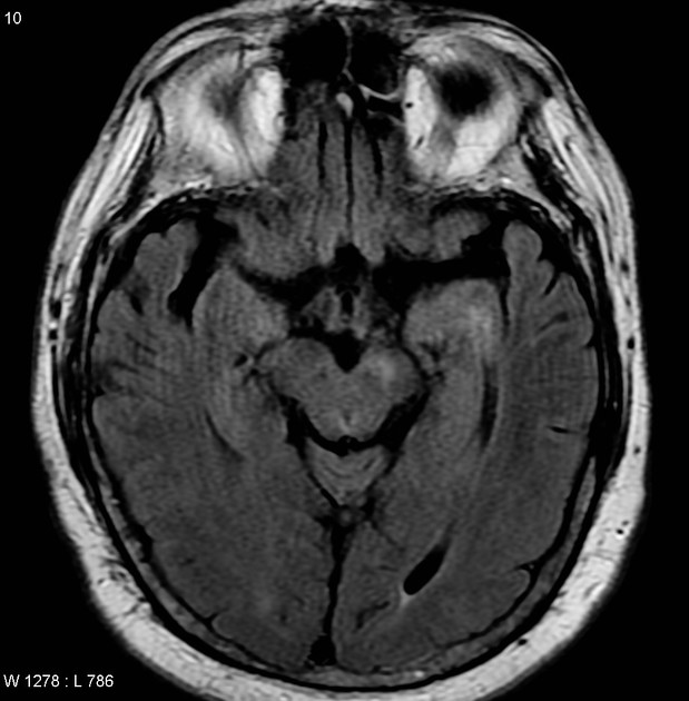
MRI of the brain demonstrates the lesion to have signal consistent with blood (high T1 and T2 centrally, peripheral hemosiderin).


Download
Info
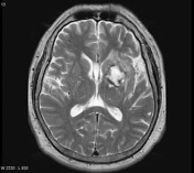
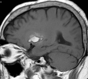
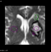
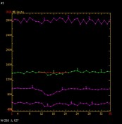
The corticospinal tracts on the left demonstrates high T2 signal in the left cerebral peduncle with reduced volume.
Case Discussion
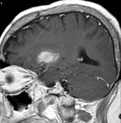
This case demonstrates how vividly a resolving hematoma can enhance.


 Unable to process the form. Check for errors and try again.