
Presentation
Painless progessive right foot drop with CT scan apparently showing no disc prolapse.
Patient Data
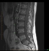


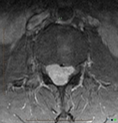
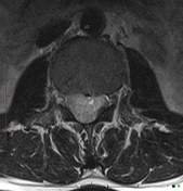
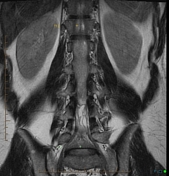

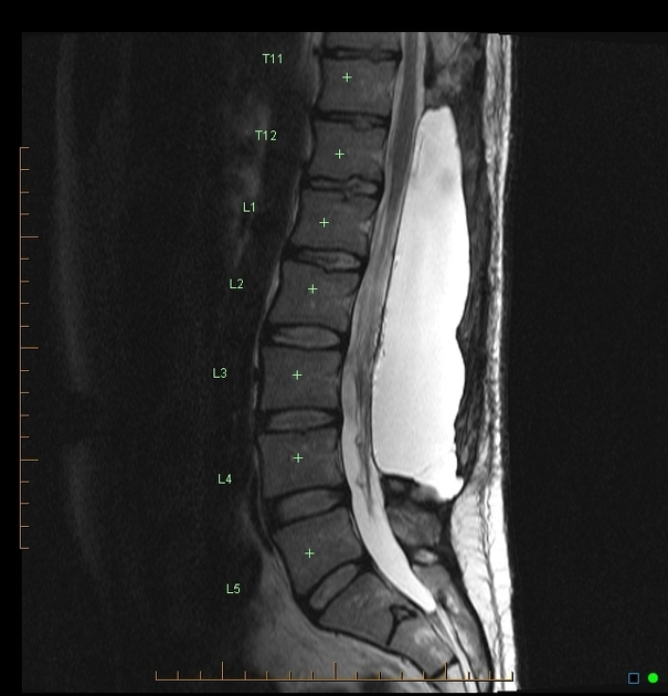
Large, enhancing extra-axial mass in the upper lumbar spinal canal deviating the conus to the left and expanding the spinal canal by way of posterior vertebral body "scalloping".
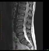
The post-op scan shows extensive laminectomy and pseudo-meningocele.
Case Discussion
Histology revealed a schwannoma. What is remarkable in this case is the relatively minor clinical signs associated with this huge mass, i.e. foot drop and no pain. This is presumably due to the slow growth of the tumor causing pressure erosion on the spinal canal. The contemporary preliminary CT (not available to me in this case) apparently did not show an obvious mass in the spinal canal. Look at the T1 sagittal images and perhaps you could make the same mistake.


 Unable to process the form. Check for errors and try again.