
Presentation
Incidental thyroid nodule.
Patient Data

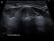


There is a 1.7 cm thyroid nodule in the isthmus showing to be well-defined, hypoechoic, and with prominent vascularization on Doppler ultrasound. The remainder of the thyroid gland (not showed) has normal appearances.
Case Discussion
The nodule was approached with FNA and came as papillary carcinoma.
Total thyroidectomy was performed:
MICROSCOPIC DESCRIPTION: Sections of thyroid isthmus show a 14mm maximum dimension lobulated tumor partially surrounded by a thick layer of fibrous tissue. The tumor is composed of papillary structures lined by atypical polyhedral cells with large irregular clear nuclei and variable amounts of pale cytoplasm. No lymphovascular or perineural invasion is identified. The tumor is clear of resection margins. The remaining thyroid parenchyma is focally fibrotic and contains a patchy lymphocytic inflammatory infiltrate. No parathyroid tissue is identified. Two lymph nodes show no evidence of malignancy.
DIAGNOSIS: Total thyroidectomy: Papillary carcinoma. - lymphovascular invasion not identified; - perineural invasion not identified; - clear of resection margins.


 Unable to process the form. Check for errors and try again.