Presentation
CSF rhinorrhea. Prior third ventriculostomy.
Patient Data
![](https://prod-images-static.radiopaedia.org/images/27774499/3a565a2c2bed1349e1aa33ec175f51_thumb.jpeg)
![This study is a stack](/packs/stack-YQKLCKBI.gif)
![](https://prod-images-static.radiopaedia.org/images/27775031/a9a50ad6859fcc6ddcbe04ef60775c_thumb.jpeg)
![This study is a stack](/packs/stack-YQKLCKBI.gif)
![](https://prod-images-static.radiopaedia.org/images/27777587/de29e2c26d31430c6ffcf749b0e0eb_thumb.jpeg)
![This study is a stack](/packs/stack-YQKLCKBI.gif)
![](https://prod-images-static.radiopaedia.org/images/27777737/3ccb27dc4000474f8fa60624da1470_thumb.jpeg)
![This study is a stack](/packs/stack-YQKLCKBI.gif)
![](https://prod-images-static.radiopaedia.org/images/27777433/46ac60283b40ccbc56be950c1b8a0b_thumb.jpeg)
![This study is a stack](/packs/stack-YQKLCKBI.gif)
![](https://prod-images-static.radiopaedia.org/images/27777688/7a13e543c60c7288c7a764dbf37e4a_thumb.jpeg)
![This study is a stack](/packs/stack-YQKLCKBI.gif)
![](https://prod-images-static.radiopaedia.org/images/27777528/3a2f39cdbdd99d6f60813b43c34899_thumb.jpeg)
![This study is a stack](/packs/stack-YQKLCKBI.gif)
![](https://prod-images-static.radiopaedia.org/images/27775451/144b29350dc291b3b853946d2a8b11_thumb.jpeg)
![This study is a stack](/packs/stack-YQKLCKBI.gif)
![](https://prod-images-static.radiopaedia.org/images/27777882/6b7f70a2ebdb1611fcb4175ffe9e25_thumb.jpeg)
![This study is a stack](/packs/stack-YQKLCKBI.gif)
![](https://prod-images-static.radiopaedia.org/images/27777807/473bc662b980d84a0826f08bfb8025_thumb.jpeg)
![This study is a stack](/packs/stack-YQKLCKBI.gif)
![](https://prod-images-static.radiopaedia.org/images/27774834/d7f20119cde21d6e0cad59e27b4ab6_thumb.jpeg)
![This study is a stack](/packs/stack-YQKLCKBI.gif)
![](https://prod-images-static.radiopaedia.org/images/27774954/4788d0ba72fb0a04770a02cc2e2ee2_thumb.jpeg)
![This study is a stack](/packs/stack-YQKLCKBI.gif)
![](https://prod-images-static.radiopaedia.org/images/27774781/f45a4fc1246178905801cd77926b3c_thumb.jpeg)
![This study is a stack](/packs/stack-YQKLCKBI.gif)
![](https://prod-images-static.radiopaedia.org/images/27775253/f94a10c33c1cda975e7265751722f4_thumb.jpeg)
![This study is a stack](/packs/stack-YQKLCKBI.gif)
![](https://prod-images-static.radiopaedia.org/images/27775340/d646016014e7d4db62f3d5e24a97ed_thumb.jpeg)
![This study is a stack](/packs/stack-YQKLCKBI.gif)
![](https://prod-images-static.radiopaedia.org/images/27774499/3a565a2c2bed1349e1aa33ec175f51_big_gallery.jpeg)
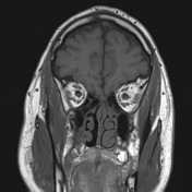
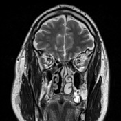
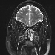
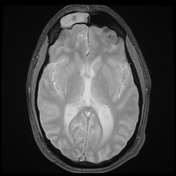
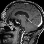
Right frontoethmoidal encephalocele with prolapse of the anterior right gyrus rectus through the floor of the right frontal sinus, through the anterior ethmoid air cells with bulging of the dural covering into the nasal cavity. The meningoencephalocele appears to contain the right orbitofrontal artery. Associated gliosis in the adjacent frontal lobe noted.
Mucosal thickening within the right frontal sinus, with underlying osteoma, and the ethmoid air cells and maxillary sinuses bilaterally. The hyperdense fluid in the frontal sinus on CT is hypointense to the mucosal thickening on T2 and relatively hyperintense on T1.
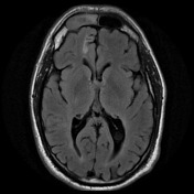
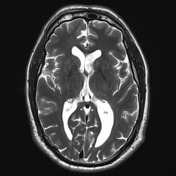
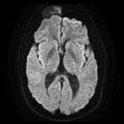
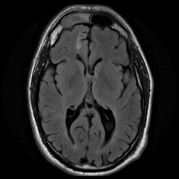
FLAIR hyperintense deep and periventricular white matter lesions are of a number greater than expected for the patient's age.
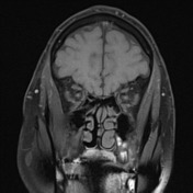
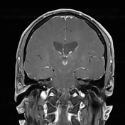
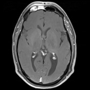
Previous mini right parietal craniotomy with underlying gliotic tract extending to the right lateral ventricle and flow void through the floor of the third ventricle in keeping with a third ventriculostomy.
Previous resection of the posterior portions of the middle and inferior turbinates.
![](https://prod-images-static.radiopaedia.org/images/27779520/99b2fbcb2f6be4df5126c3635e0fb6_thumb.jpeg)
![This study is a stack](/packs/stack-YQKLCKBI.gif)
![](https://prod-images-static.radiopaedia.org/images/27779552/291bdee547984ddb014b5e4a6a2a20_thumb.jpeg)
![This study is a stack](/packs/stack-YQKLCKBI.gif)
![](https://prod-images-static.radiopaedia.org/images/27779520/99b2fbcb2f6be4df5126c3635e0fb6_big_gallery.jpeg)
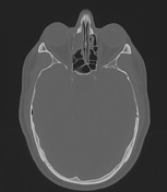
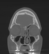
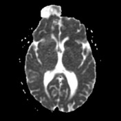
CT demonstrates a defect of the right paramedian skull base just to the lateral margin of the olfactory groove. Much of the right frontal sinus is opacified as is the anterior ethmoid air cells and the medial margin of the middle ethmoid air cells on the right. These findings are likely to represent a combination of the opacification and sinus mucosal thickening associated with the meningoencephalocoele.
Case Discussion
The patient went on to have surgery with resection of the encephalocele and repair of the base of skull.
Histology:
The section shows fragments of inflamed sinus mucosa, viable unremarkable bone and a fragment of brain parenchyma which has distorted architecture and shows reactive astrocytic gliosis. The features in this fragment are consistent with an encephalocele. No evidence of tumor is seen.
FINAL DIAGNOSIS: Features consistent with encephalocele.