
Presentation
Headache
Patient Data


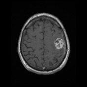
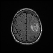
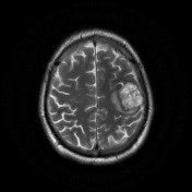

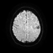

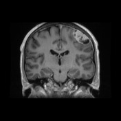
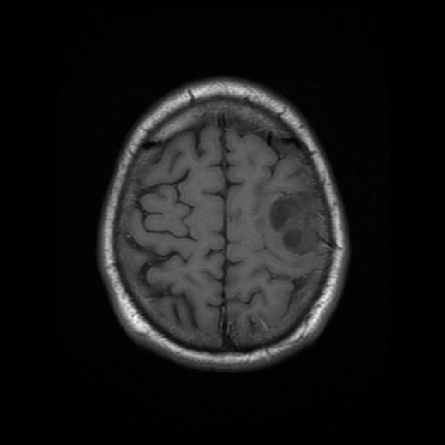
A lobulated extra-axial mass is present indenting the left frontal lobe. It is of high T2 signal and demonstrates vivid enhancement except for areas of cystic change.
Case Discussion
The patient went on to have surgery.
Histology
MICROSCOPIC DESCRIPTION: Sections show a dural-based tumor composed of variably sized thin-walled blood vessels, microcysts and intervening stroma containing tumor cells with abundant eosinophilic cytoplasm, round to oval nuclei with fine chromatin and inconspicuous nucleoli. No necrosis or mitoses are seen. No brain invasion is identified. There is no evidence of malignancy.
Immunohistochemical results show tumor cells stain: EMA weak+, PR+, Inhibin- and Topoisomerase 5-8%.
FINAL DIAGNOSIS: microcystic meningioma (WHO Grade I).


 Unable to process the form. Check for errors and try again.