
Presentation
Pelvic pain in a patient with known uterine didelphys.
Patient Data
Age: 25 years
Gender: Female
From the case:
Uterine didelphys and tubo-ovarian abscess


Download
Info
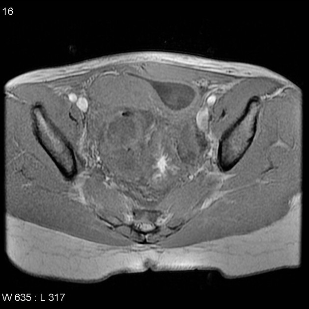
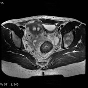
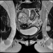
Uterine didelphys is noted.
A large right adnexal complex lesion with dilated right Fallopian tube filled with fluid. Tiny low signal intensity foci in the non-dependent portions consistent with complicated gas formation. Minimal perifocal free fluid is seen.
Case Discussion
Female patient with uterine didelphys. The right cervix was seen at colonoscopy to end in the rectum and subsequent tubo-ovarian abscess formed.
Although surgical intervention was planned, the wait list was long and the collection was subsequently drained via a transvaginal approach.


 Unable to process the form. Check for errors and try again.