
Celoria-Patton classification of interrupted aortic arch (illustration)
Diagnosis not applicable

Download
Info

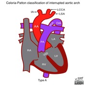
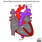
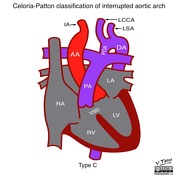
This diagram depicts the types of interrupted aortic arch according to the site of interruption:
- type A: distal to the left subclavian artery (LSA) - 2nd most common
- type B: between the left common carotid artery (LCCA) and left subclavian artery (LSA) - most common
- type C: between the innominate artery (IA) and left common carotid artery (LCCA)
(AA - ascending aorta; DA - descending aorta; PDA - patent ductus arteriosus; VSD - ventricular septal defect; PA - pulmonary artery; RA - right atrium; LA - left atrium; RV - right ventricle; LV - left ventricle)


 Unable to process the form. Check for errors and try again.