
Presentation
35-year-old male patient with congenital blindness and recurrent seizures (myoclonic type).
Patient Data
Age: 35 years
Gender: Male
From the case:
Septo-optic dysplasia with schizencephaly


Download
Info

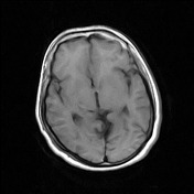
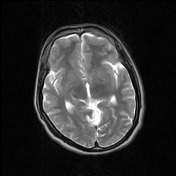
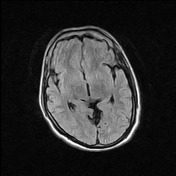
Selected MR images show nipple like ependymal projection in the region of the left lateral ventricle body (parietal region) which is lined by grey matter signal reaching up to the cortical surface on axial T2WI/FLAIR. Coronal T1WI are also showing this abnormality.
T2 axial and FLAIR axial and T1 coronal images demonstrated thinned optic nerves.
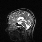
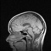
Sagittal T1WI/T2WI images are showing hypoplastic rostrum/genu of corpus callosum
Case Discussion
Patients clinical history coupled with MR findings are suggestive of closed lip schizencephaly with septo-optic dysplasia and possible dysgenesis of the corpus callosum.


 Unable to process the form. Check for errors and try again.