
Presentation
Dysphagia and vomiting
Patient Data
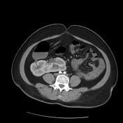

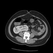
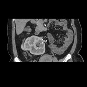
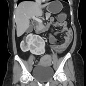


Arrested ascent of right kidney with superior border reaching only the L2 vertebra. Ectopic left kidney is seen to the right of midline and fused to the medial aspect of right kidney. Malrotated right kidney with hilum pointing anterolaterally.
Both kidneys have an extrarenal pelvis. Normal left ureter insertion.
Origin of renal vessels from abdominal aorta is beneath the inferior mesenteric artery with left kidney receiving dual arterial supply - renal branch and right common iliac arterial branch.
Case Discussion
This patient came for low back ache evaluation under MRI and the absence of a right kidney in it's normal position, accompanied by an empty left renal fossa, prompted further evaluation. Whilst the CT abdomen was done for different purposes all together, this anatomic variant was confirmed.


 Unable to process the form. Check for errors and try again.