
Presentation
An incidental finding in a case recently diagnosed with left breast cancer for staging work up.
Patient Data
Age: 60 years
Gender: Female
From the case:
Giant hepatic hemangioma


Download
Info
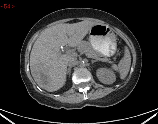
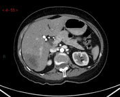
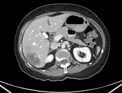
Large hepatic lesion, involving predominantly segments VI and VII, with peripheral nodular discontinuous enhancement with progressive centripetal fill-in; likely representing a giant hemangioma rather than metastasis.
Linear hypodense filling defect in the left renal vein consistent with a thrombus.
Case Discussion
An incidentally noted giant hemangioma in the liver is an example of an atypical hepatic hemangioma.
Giant hepatic hemangiomas (or "giant hepatic slow flow venous malformations") are defined to be hemangiomas >4 cm.


 Unable to process the form. Check for errors and try again.