
Presentation
Left side conductive hearing loss, left retroauricular swelling.
Patient Data
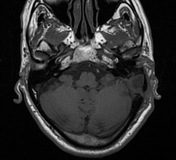

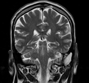
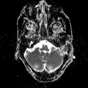
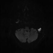
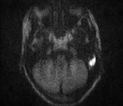
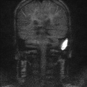

Evidence of left mastoid lesion measuring 1.2 x 3 cm expressing T1 hypointense signal and T2 bright signal. No post-gadolinium enhancement. It showed marked diffusion restriction, best seen on HASTE diffusion-weighted (non echo-planar) sequence.
It shows overlying 9 mm calvarial defect at the occipitomastoid suture and wide destruction of the sigmoid plate. There is thick enhancing granulation tissue at the interface with the sigmoid sinus posteriorly. No labyrinthine or other intracranial lesions.
Loss of signal void on coronal T2 images of the left sigmoid and distal left transverse sinuses suggestive of sigmoid thrombophlebitis.
Case Discussion
This case shows MRI imaging features of left mastoid cholesteatoma with surrounding inflammatory granulation tissue showing marked diffusion restriction and bone destruction. This is complicated by sigmoid thrombophlebitis and temporal bone erosion.
MRI with diffusion is recommended for all cases with suspected cholesteatoma on CT for confirmation and detection of possible complications, preferably HASTE diffusion-weighted (non echo-planar) sequence. Choleateatoma shows marked diffusion restriction with low ADC values.
Complications include sigmoid sinus thrombosis, bone erosions (best by CT), meningitis, brain abscess, facial paralysis, infective cochleolabyrinthitis.


 Unable to process the form. Check for errors and try again.