
Presentation
Fatigue and muscle weakness for 6 months. Blood tests showed hypercalcemia and elevated levels of PTH, raising the suspicion of primary hyperparathyroidism.
Patient Data


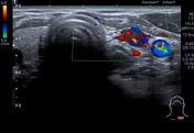

Immediately caudal to the left thyroid lobe there is a 5 mm rounded structure, slightly hypoechoic compared to the thyroid tissue. Vivid Doppler signal in the lesion, suggesting rich vascularization. Parathyroid adenoma is likely.
Due to this finding, a Tc99m-sestamibi SPECT/CT was performed.


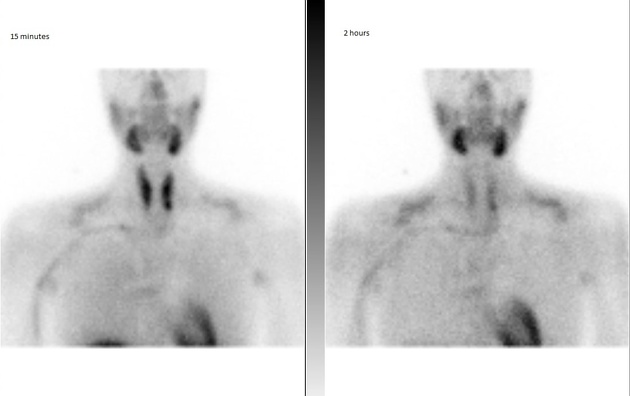
Immediately caudal to the left thyroid lobe, there is punctate uptake in the early phase (15 min registration) that remains active during the late phase (2 hours). The localization of this uptake matches the lesion identified with ultrasound, thus proving it to be a parathyroid adenoma.
Case Discussion
This case serves to demonstrate the typical ultrasound appearance of a parathyroid adenoma - a rounded, slightly hypoechoic lesion with increased vascularity. However, ultrasound characteristics may vary considerably.
Tc99m-sestamibi scans are often used in combination with ultrasound, either to verify that a lesion detected with ultrasound indeed is a parathyroid adenoma, or to increase diagnostic accuracy in cases of a negative ultrasound. This often provides important information to the surgeon, especially in patients with ectopic parathyroid glands in locations inaccessible by ultrasound.
It is important to recognize the diagnostic overlap of these two methods, the combination of which offers a preoperative diagnostic sensitivity of 95% and specificity of 91% according to one study 2.


 Unable to process the form. Check for errors and try again.