
Patient Data
Age: Adult
- Note: This case has been tagged as "legacy" as it no longer meets image preparation and/or other case publication guidelines.
From the case:
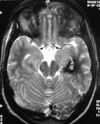
Cerebral cavernoma

Download
Info
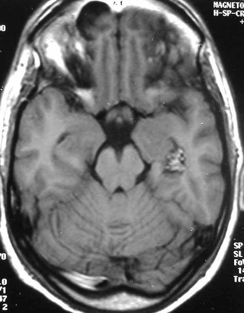
Well-defined mass in left medial temporal lobe having a mulberry-like appearance suggestive of a cavernoma. Note marked hemosiderin staining on T2WI.


 Unable to process the form. Check for errors and try again.