
Presentation
Right knee trauma, pain and swelling
Patient Data


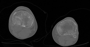
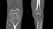
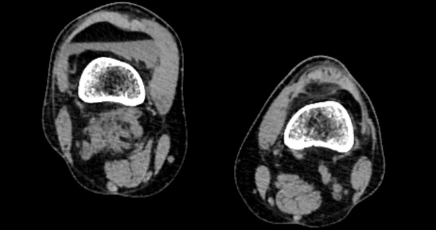
Comminuted fracture right lateral tibial condyle reaching the articular surface and extending to the proximal tibial shaft. Abundant right knee effusion with fat-blood level consistent with lipohemarthrosis. Normal osseous features of the distal aspect of the right femur and proximal part of the right fibula. Normal CT features of the left knee joint.
Case Discussion
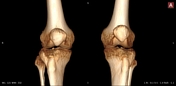
This case represents a Schatzker I lateral tibial plateau fracture seen as a wedge-shaped pure cleavage fracture of the lateral tibial plateau with no depression. The fracture should be examined in at least two planes and in bone window setting as undisplaced fractures can be easily missed. Also, undisplaced ungapping fractures hardly appear at 3D images. This is associated with moderate right knee effusion showing a dependent fat-blood level. Lipohemarthrosis occurs with fractures of long bones which involve the medullary cavity, which leads to leakage of fat from the bone marrow, with intra-articular extension of the fracture lines. Knee joint fractures are the commonest cause of lipohemarthrosis, however, it can occur at the hip, shoulder or elbow joints. It can be easily seen in horizontal beam plain x-ray, as well as CT/MRI due to a marked density difference between fat and blood with fluid levelling, called the FBI sign.


 Unable to process the form. Check for errors and try again.