Presentation
Pathologically-proven brown tumor of the ulna.
Patient Data
Age: 65 years
Gender: Female
From the case:
Parathyroid adenoma
![](https://prod-images-static.radiopaedia.org/images/31977692/0ac4dd3e84c273e48ded8c48390661_thumb.jpeg)
![This study is a stack](/packs/stack-YQKLCKBI.gif)
![](https://prod-images-static.radiopaedia.org/images/31978280/2659d1e67184699138f06ac1688b99_thumb.jpeg)
![This study is a stack](/packs/stack-YQKLCKBI.gif)
![](https://prod-images-static.radiopaedia.org/images/31978757/6ce828da3a240cca3dc667b87c6692_thumb.jpeg)
![This study is a stack](/packs/stack-YQKLCKBI.gif)
![](https://prod-images-static.radiopaedia.org/images/31977179/dffe9b67e1f49a03c671669a6fd53e_thumb.jpeg)
![This study is a stack](/packs/stack-YQKLCKBI.gif)
![](https://prod-images-static.radiopaedia.org/images/31978512/fc028f832f2e61b30abfe06d58ccd3_thumb.jpeg)
![This study is a stack](/packs/stack-YQKLCKBI.gif)
![](https://prod-images-static.radiopaedia.org/images/31977981/b63c82330d5857a4a0fce9851c978b_thumb.jpeg)
![This study is a stack](/packs/stack-YQKLCKBI.gif)
Download
Info
![](https://prod-images-static.radiopaedia.org/images/31977692/0ac4dd3e84c273e48ded8c48390661_big_gallery.jpeg)
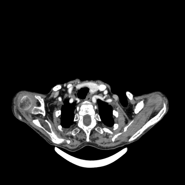
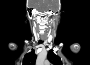
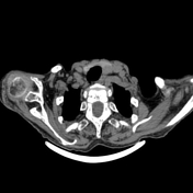
A well-defined lesion seen at the inferior border of the left thyroid lobe, with reduced enhancement compared to the normal thyroid gland in the arterial phase but with greater washout than the thyroid gland in the delayed phase. The lesion measured about 16 x 12 mm with no calcification.
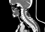
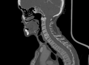
Diffuse decreased density of the imaged skeleton.
Case Discussion
The patient had a pathologically proven brown tumor of the ulna. A parathyroid adenoma was suspected as the underlying cause and was confirmed by CT.