
Presentation
Mild cognitive impairment with headache. Evaluate for intracranial abnormality.
Patient Data


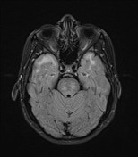
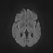
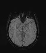
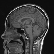
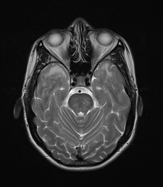
Multiple supratentorial T2/FLAIR hyperintense foci involving the subcortical and periventricular white matter bilaterally. There is a bitemporal symmetric predominance. It is difficult to comment on the juxtacortical (U-fibers) on these images, although we suspect that the involvement is primarily that of subcortical white matter. There is no restricted diffusion and no associated significant microhemorrhage.
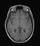
There is no significant brain atrophy.
Case Discussion
The findings were, clinically and radiologically, in favor of CADASIL. The bitemporal distribution is classic of this entity. The main differential diagnosis would be MELAS and CNS vasculitis.


 Unable to process the form. Check for errors and try again.