
Presentation
Two days of increasing headaches, now right sided weakness.
Patient Data

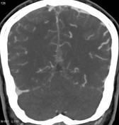
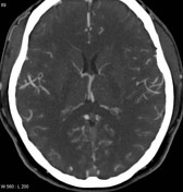
This non-contrast CT scan of the brain demonstrates hyperdense superior sagittal sinus, straight sinus, vein of Galen, transverse sinus on the left and a number of cortical veins. In addition a region of low density with foci of hemorrhage is noted in the partial lobe on the right.


Selected axial, coronal and sagittal images from a CT venogram confirms extensive venous thrombosis as demonstrated on the non-contrast CT.
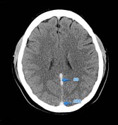

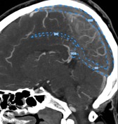


Thrombosed hyperdense superior sagittal sinus (SSS), straight sinus (SS), vein of Galen (VOG), transverse sinus (TS) on the left and a number of cortical veins (CV). In addition the inferior sagittal sinus (ISS) is not visualized on CT venogram and is presumably also thrombosed (or hypoplastic). A region of low density with foci of hemorrhage (yellow dotted line) is noted in the partial lobe on the right consistent with a venous infarct.
Case Discussion
This young woman spontaneously developed extensive dural venous sinus thrombosis with right sided hemorrhage. The only risk factor identified was the oral contraceptive pill.


 Unable to process the form. Check for errors and try again.