Presentation
Presented with 13 wks 6 days amenorrhea without any complaints. Previous failed early pregnancy about 10 months back.
Patient Data

Single live intrauterine fetus shows a cystic lesion involving posterior cervical region. It shows extension to thoracic region. There are multiple septa within the lesion. Rest of the fetal anatomical survey was normal for maturity which corresponded to the period of amenorrhea.

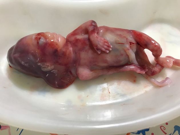
Abortus shows redundant soft tissue in the cervical region.
Case Discussion
Ultrasound findings favor cystic hygroma. Later, chorionic villus sampling was done by fetal medicine consultant. Karyotyping revealed trisomy 21.
Abortus photo: Courtesy treating obstetric surgeon Dr. Drashti R. Patel.


 Unable to process the form. Check for errors and try again.