
Presentation
Known treated renal cell carcinoma patient with headache and intermittent fever
Patient Data


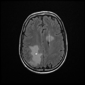
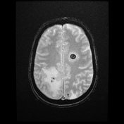
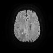
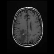
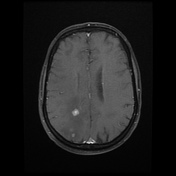
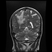
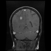
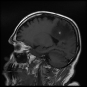
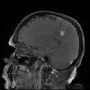
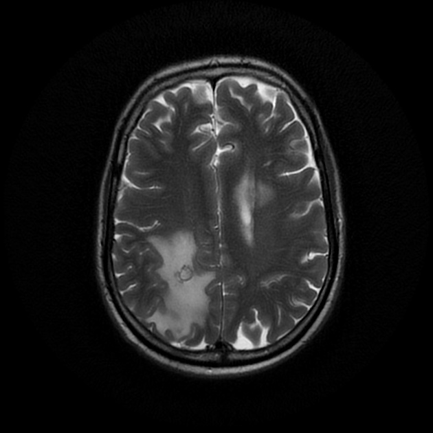
Intra axial grey white matter junction mass lesions seen in left frontal and right parietal lobes. Lesions are T1 hypointense, T2 and FLAIR hyperintense, mild diffusion restricted and showing intense gadolinium enhancement. Areas of T1 hyperintensity with blooming on T2* also seen suggestive of hemorrhage. Moderate vasogenic edema seen in right parietal lobe around the lesion causing mild effacement of ipsilateral lateral ventricle. No brain stem or cerebellar lesions.
Case Discussion
Hemorrhagic hypervascular metastases are typical of few organs - kidneys, thyroid, neuroendocrine tumors. These are typical imaging findings of brain metastases in a known renal cell carcinoma patient.


 Unable to process the form. Check for errors and try again.