
Presentation
Incidental hyperechogenic lesions on ultrasound, for further imaging characterization.
Patient Data
Age: 50-years
Gender: Female


Download
Info
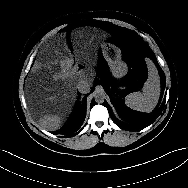
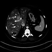
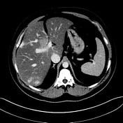
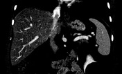
The liver is of lower attenuation than normal, consistent with diffuse steatosis with a region of focal fatty sparing deep in segment 4. Multiple liver lesions are again demonstrated and these have a typical enhancement pattern of hemangiomata. There is also a 7 cm left lower pole renal simple cyst. No significant abnormality in the spleen, pancreas, either adrenal gland or right kidney. No upper abdominal para-aortic lymphadenopathy.
Case Discussion
This dedicated liver CT demonstrates diffuse steatosis and multiple liver mass like lesions consistent with hemangiomas (hemangiomata).


 Unable to process the form. Check for errors and try again.