
Presentation
Seizure.
Patient Data

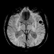
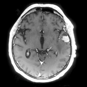
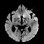
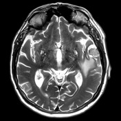
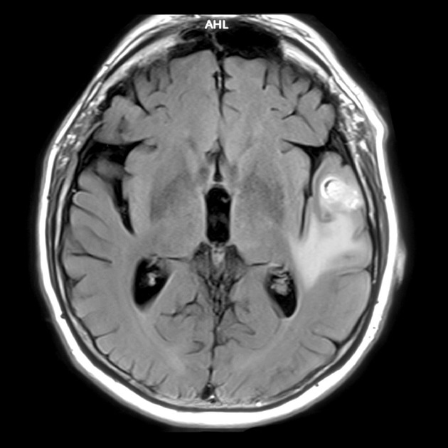
Selected MRI images demonstrate an extra-axial mass on the right with edema within the right temporal lobe. The mass demonstrates signal loss on SWI and vivid contrast enhancement. Non-contrast T1 (not shown) demonstrates some intrinsic high T1 signal.
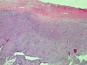
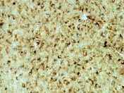

The sections show a moderately hypercellular tumor. This consists of epithelioid and spindle cells which are heavily laden with globular brown pigment and show strong immunostaining for the melanocyte markers, melan-A.
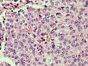
Tumor cells show moderate nuclear pleomophism with vesicular nuclei containing conspicuous nucleoli.
Focal dural attachment is identified.
DIAGNOSIS: Melanocytic tumor with features favoring meningeal melanocytoma.
Case Discussion
Meningeal melanocytomas are rare tumors and it is difficult to make the diagnosis preoperatively. The differential includes other melanotic lesions (e.g. melanotic meningioma and intracranial metastatic melanoma) and hemorrhagic lesions (e.g. hemangioblastoma hemorrhagic metastases).


 Unable to process the form. Check for errors and try again.