
Presentation
Recurrent shoulder dislocation
Patient Data
Age: 35 years
Gender: Male
From the case:
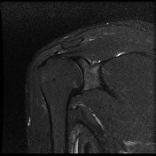
Bankart lesion

Download
Info

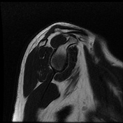
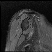
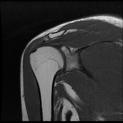
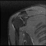
Intermediate signal intensity seen in all pulse sequences in the supraspinatus tendon. PD high signal intensity seen in anteroinferior labrum from 3 to 6 o clock.
From the case:
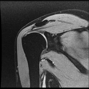
Bankart lesion
Download
Info

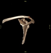
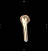
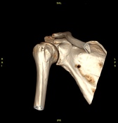
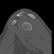
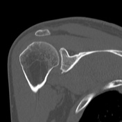
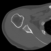
Bony depressed 7 mm fracture seen in the anteroinferior glenoid margin. Osteodegenerative changes seen in the posterosuperolateral humeral head.
Case Discussion
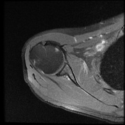
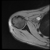
Recurrent anterior dislocation with a torn anteroinferior labrum and associated glenoid bony fracture is a typical case of bony Bankart. There is associated Hill Sach lesion in the posterosuperolateral humeral head. <5% glenoid volume loss in this case.
This is a typical example of bony Bankart with Hill Sach lesion.


 Unable to process the form. Check for errors and try again.