
Presentation
Abdominal distension and pain after VP shunt insertion
Patient Data
Age: 8 years
Gender: Female
From the case:
Peritoneal/subcapsular CSF pseudocyst


Download
Info
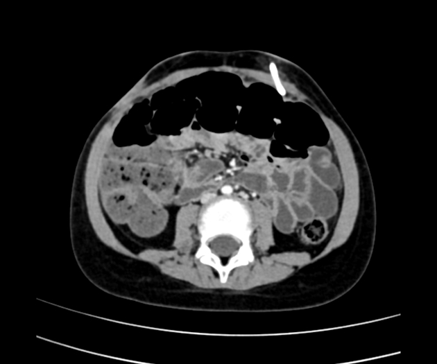
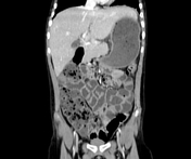
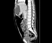
VP shunt is seen inserting left side anterior abdominal wall just above the level of umbilicus.
A large water density collection in the epigastric region anterior to left lobe of liver engulfing the VP shunt and exerting mass effect over adjacent liver parenchyma.
Case Discussion
The features are considered as peritoneal CSF pseudocyst, however it is possible that the VP shunt has penetrated the hepatic capsule and the collection can be subcapsular in fact.


 Unable to process the form. Check for errors and try again.