
Presentation
Awoke with right sided weakness and dysphasia. Background of non-small cell lung cancer, being treated with chemotherapy and radiotherapy.
Patient Data
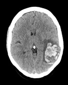


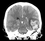
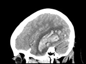

There are confluent hemorrhagic foci within the left temporal lobe in a peripheral subcortical distribution associated with surrounding edema. The distribution is atypical for an infarct involving the left middle cerebral artery territory. Note is made of a filling defect within the torcula Herophili (empty delta sign) and hyperdense left transverse dural venous sinus.
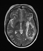
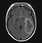
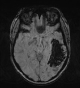
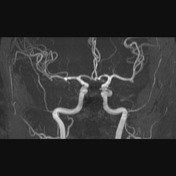
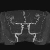
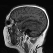
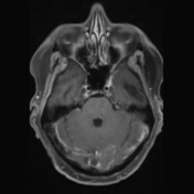
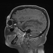
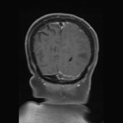
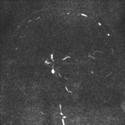
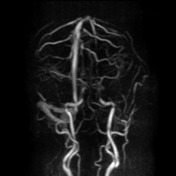

MRI confirms the venous hemorrhagic infarct within the left temporal lobe and thrombosis within the left transverse and sigmoid dural venous sinuses.
Case Discussion
This is a case that should prompt you to consider venous infarction upon reviewing the first non-contrast CT imaging. The left temporal lobe hemorrhagic infarction has a gyriform pattern within a non-arterial location/territory.


 Unable to process the form. Check for errors and try again.