
Presentation
Four years history of progressive rigidity, choreoathetosis, and dementia.
Patient Data


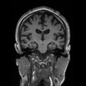

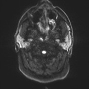
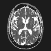
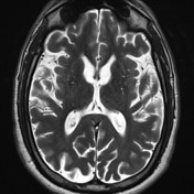
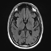
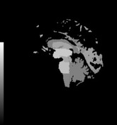
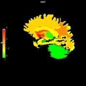
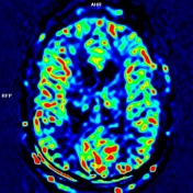
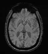
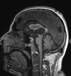
There is diffuse brain volume loss involving both grey and white matter along the cerebral hemispheres, including marked bilateral caudate and putaminal atrophy. The frontal horn width to intercaudate distance ratio (FH/CC) is reduced (ratio=1.51 - normal range 2.2 - 2.6) and the intercaudate distance to inner table width ratio (CC/IT) is increased (ratio=0.23 - normal range 0.09 - 0.12). A few white matter FLAIR hyperintensities are nonspecific and within the normal range for the patient's age group. No evidence of restricted effusion or abnormal susceptibility artefacts. ASL perfusion is unremarkable.
Case Discussion
This patient has a known family history and a recent diagnosis with a positive gene for Huntington's disease. MRI shows supportive imaging features characterized by brain volume loss with morphological changes of Huntington's disease: caudate nuclei atrophy.


 Unable to process the form. Check for errors and try again.