
Presentation
Neck pain.
Patient Data
Age: 30 years
Gender: Male
From the case:
Cervical schwannoma


Download
Info
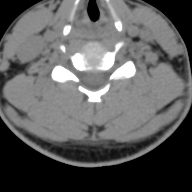
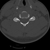
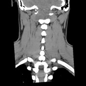
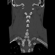
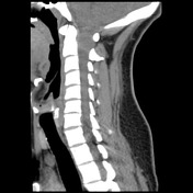
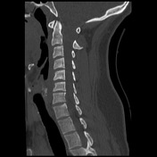
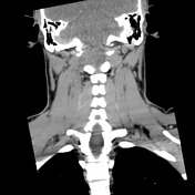
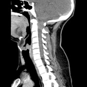
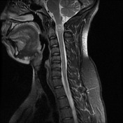
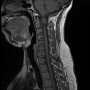
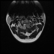
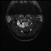
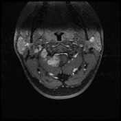
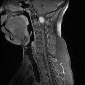
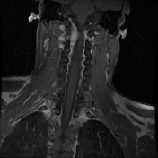
Large mass centered on the right C1/2 neural exit foramen with associated foraminal widening and bony erosion.
From the case:
Cervical schwannoma

Download
Info
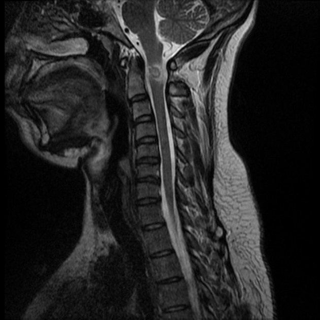
Large mass centered on the right C1/2 neural exit foramen with associated foraminal widening and bony erosion. The mass has a "dumb bell" morphology and is isointense to brain on T1WI and T2WI with vivid post contrast enhancement. The spinal cord is displaced and compressed to the left.
Case Discussion
The patient went on to surgical resection, and schwannoma was confirmed as the diagnosis on histopathology.


 Unable to process the form. Check for errors and try again.