
Presentation
Follow-up in melanoma
Patient Data
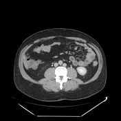

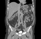
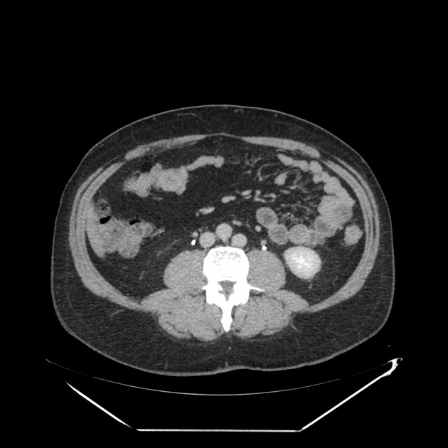
The CT examination documented a complete duplication of the lower vena cava.
Case Discussion
Although this abnormality is not very frequent, it must be correctly recognized above all for the implications that it can have. For example, of the positioning of a caval filter in case of risk of pulmonary embolism, or it could influence the correct diagnostic approach in the treatment of portal hypertension where the need arises to create a port-systemic shunt or, again, in the management of kidney transplantation.
This condition should not be confused with the transposition of the IVC, in which the lower caval vein lies to the left of the aorta.
Other anomalies concern the position of the left renal vein, the most frequent of which is represented by its retroaortic course.


 Unable to process the form. Check for errors and try again.