Presentation
Workup for hypogonadotropic hypogonadism. Right eye congenital blindness.
Patient Data
![](https://prod-images-static.radiopaedia.org/images/36260642/8c8e6162839ced0a3c9b9a689b2609_thumb.jpeg)
![This study is a stack](/packs/stack-YQKLCKBI.gif)
![](https://prod-images-static.radiopaedia.org/images/36260739/07330ecfc611d87771b68da44bf095_thumb.jpeg)
![This study is a stack](/packs/stack-YQKLCKBI.gif)
![](https://prod-images-static.radiopaedia.org/images/36260938/810e500c79096dfd3fad55fb899cf1_thumb.jpeg)
![This study is a stack](/packs/stack-YQKLCKBI.gif)
![](https://prod-images-static.radiopaedia.org/images/36261028/a65d96efc69b9181b7236e9b75a923_thumb.jpeg)
![This study is a stack](/packs/stack-YQKLCKBI.gif)
![](https://prod-images-static.radiopaedia.org/images/36261215/f9a11a70c0f2f22c01a7e7ce188a3b_thumb.jpeg)
![This study is a stack](/packs/stack-YQKLCKBI.gif)
![](https://prod-images-static.radiopaedia.org/images/36260642/8c8e6162839ced0a3c9b9a689b2609_big_gallery.jpeg)
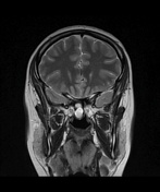
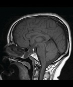
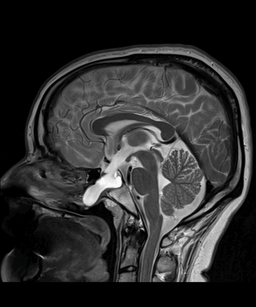
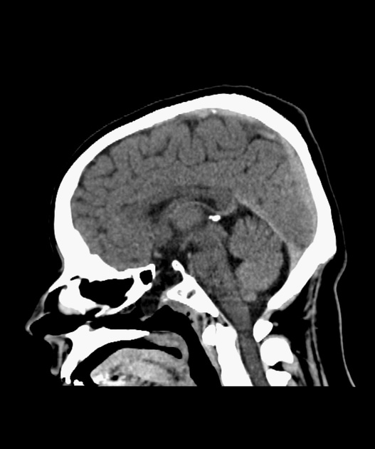
Transsphenoidal meningocele anterior to the dorsum sella projecting into the nasopharynx through a large craniopharyngeal canal. There is a communication between the anterior recess of the third ventricle and the meningocele. The pituitary stalk and gland can't be identified. There is a thin layer of contrast enhancing tissue at the bottom of the meningocele and a small nodular contrast enhancing tissue at the tip of the meningocele, possibly containing pituitary tissue.
Additionally, abnormal morphology of the right eye globe is noted.
![](https://prod-images-static.radiopaedia.org/images/36266620/efdf36f7fad640377c1f3c1b37a845_thumb.jpeg)
![This study is a stack](/packs/stack-YQKLCKBI.gif)
![](https://prod-images-static.radiopaedia.org/images/36266839/bae696d0ab2e4101d9d7552d2dbfa8_thumb.jpeg)
![This study is a stack](/packs/stack-YQKLCKBI.gif)
![](https://prod-images-static.radiopaedia.org/images/36267102/90f01b1a5845074c8e5b48a1d8c033_thumb.jpeg)
![This study is a stack](/packs/stack-YQKLCKBI.gif)
![](https://prod-images-static.radiopaedia.org/images/36395444/d60080ee1a73350ff97b5118d52797_thumb.jpeg)
![This study is a stack](/packs/stack-YQKLCKBI.gif)
![](https://prod-images-static.radiopaedia.org/images/36395564/e4820a48e46efdf52728bed801998c_thumb.jpeg)
![This study is a stack](/packs/stack-YQKLCKBI.gif)
![](https://prod-images-static.radiopaedia.org/images/36395751/89dd59c5e2252f531d8dcfa0a950e3_thumb.jpeg)
![This study is a stack](/packs/stack-YQKLCKBI.gif)
![](https://prod-images-static.radiopaedia.org/images/36266620/efdf36f7fad640377c1f3c1b37a845_big_gallery.jpeg)
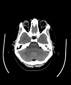
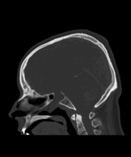
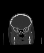
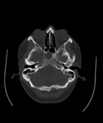
CT-examination with the same findings. Additionally, incomplete cleft palate is noted (best appreciated with axial bone window images).
Case Discussion
This is a case of congenital transsphenoidal meningocele presenting with hormonal disturbances. Eye globe malformations, as this patient had, have been described in cases with transsphenoidal meningocele. The patient went on to have transsphenoidal surgery.