
Presentation
Knee pain and swelling of 2 years duration. History of knee trauma 5 years ago.
Patient Data


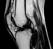
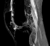
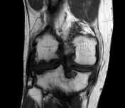
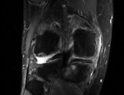
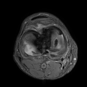
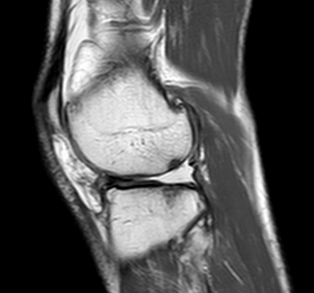
The posterior horn of the lateral meniscus shows a radial tear with a wide gap between the torn edges measuring 13 mm in length and giving the appearance of a ghost meniscus on sagittal cuts. This gaping allows contact between the lateral femoral and tibial condyles which results in cartilage denudation and osteoarthritis.
Moreover, there is longitudinal horizontal tear of posterior horn of medial meniscus interrupting the inferior articular surface, ACL injury, moderate effusion, and baker cyst.


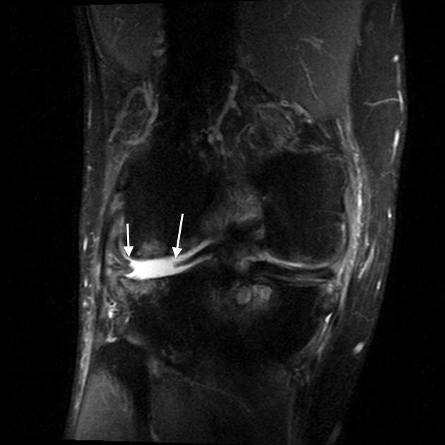
A gap is formed between the torn edges (white arrows on coronal) and black arrows on axial images.
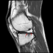
Focal cartilage loss and subchondral degeneration are noted on sagittal image (between red arrows).
Case Discussion
Radial tears occur due to shear forces and start at the inner free margin of the meniscus extending toward the periphery. They disrupt the longitudinal collagen fibers of the meniscus and when reaching the peripheral margin can result in gap between the torn edges. This allows contact between the articular surfaces and later on development of osteoarthritis.


 Unable to process the form. Check for errors and try again.