
Presentation
Right loin pain.
Patient Data
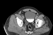


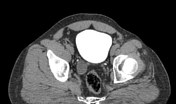

The right kidney shows markedly dilated upper calyx reaching 8 cm in maximum AP diameter, with thinned overlying renal parenchyma. It connects to a non-opacified upper moiety ureter which is moderately dilated along its whole length, until it inserts at the medial aspect of the prostatic urethra.The lower calyx is normal in size draining into the lower moiety which is seen inserting at the normal vesicoureteric junction. Otherwise, the right kidney is normal in size and position. No renal stone, expanding lesion identified.
The left kidney is normal in size, shape, and position. No renal stone, expanding lesion, or back pressure changes identified.
Normal contour of the urinary bladder, with uniform wall and no filling defects noted.
Case Discussion
Ectopic insertion of the ureter is rare urinary tract anomaly, inserting into nearby pelvic organs such as the seminal vesicle, prostatic urethra, or vagina. It is usually associated with duplex kidney anomaly. According to the Weigert-Meyer law, the upper pole moiety ureter drains infero-medial to the normal lower moiety ureter, and so it is more likely for ectopic ureteral insertion.
Here, this case demonstrates duplex kidney with double ureter and ectopic insertion of the right upper moiety ureter into the prostatic urethra. There is moderate dilatation of the upper moiety of the right kidney and its ureter, and secondary atrophic changes of the right renal upper pole.
In males, the prostatic urethra is the commonest site of ectopic insertion, accounting for ~50%. Delayed contrast excretory phase in CT urography is the most sensitive, economic, and readily available test for diagnosing ectopic ureters 1.


 Unable to process the form. Check for errors and try again.