
Presentation
History of myocardial infarction, coronary stenting (LAD, OM, R-PLB) .
Patient Data


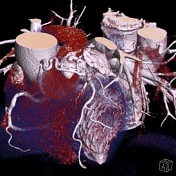
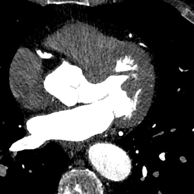
Axial CT, VRT images show a tortuous vessel arising from the right coronary artery, which passes anterior to the pulmonary artery and forms a network before it enters the main pulmonary artery.
Findings are consistent with a coronary artery fistula (CAF).
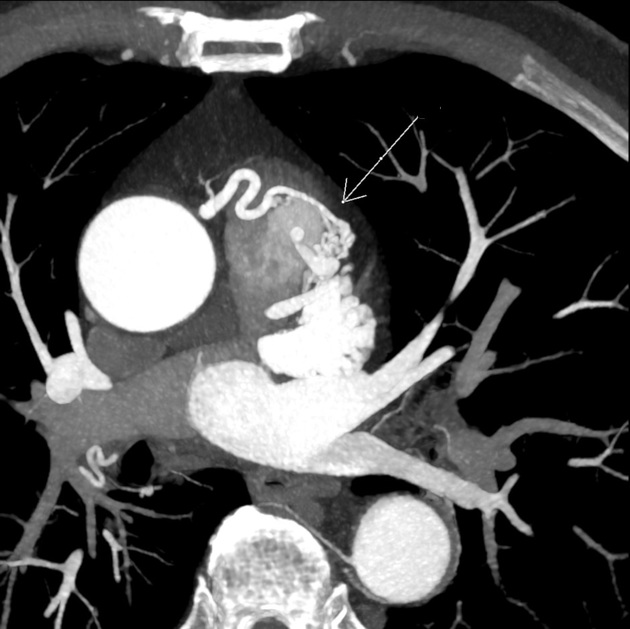
Coronary arteriovenous fistula (white arrow) on axial arterial phase MIP CT. The MIP image nicely shows diffusion of the resulting contrast blush within the pulmonary artery.
Case Discussion
This findings are noticed incidentally at CT cardiac imaging for coronary stents visualization.
In this case surgical correction was not indicated due to asymptomatic clinical presentation and elderly age.
CT follow-up was recommended.


 Unable to process the form. Check for errors and try again.