
Presentation
Visual disturbance.
Patient Data


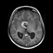
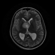
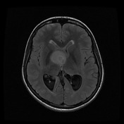
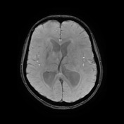
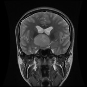
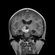

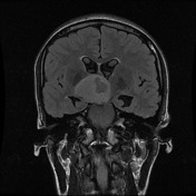
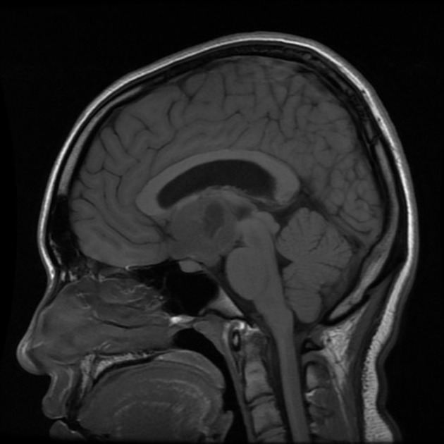
A sizable heterogeneously enhancing mass is centered on the hypothalamus and chiasm with some hydrocephalus. Features, especially in this age group, are of a hypothalamic-optochiasmatic glioma (pilocytic or pilomyxoid astrocytoma).
MICROSCOPIC DESCRIPTION:
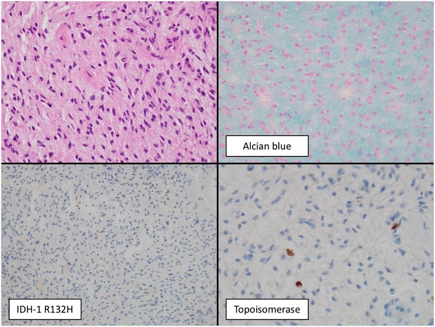
An H&E stained section demonstrates a moderately cellular tumor composed of elongated oval to wavy nuclei set within a fibrillary and myxoid stroma. There are scattered thin-walled blood vessels present. No necrosis or microvascular proliferation is seen. There are no mitoses identified. The Alcian blue stain confirms myxomatous tumor stroma. (Histology courtesy of Prof. Michael Gonzales)
Immunohistochemistry results show tumor cells stain:
- IDHl - negative.
- ATRX - positive.
- P53 - negative.
- H3K27M - negative.
- H3K27ME3 - positive.
- Topoisomerase proliferation index - 2-3%
FINAL DIAGNOSIS: Pilomyxoid astrocytoma (WHO grade I).
Case Discussion
Pilomyxoid astrocytomas and pilocytic astrocytomas are related but believed to be distinct. It is difficult to distinguish between them on imaging.


 Unable to process the form. Check for errors and try again.