
Cerebral autosomal dominant arteriopathy with subcortical infarcts and leukoencephalopathy - CADASIL
Presentation
History of recurrent transient ischemic attacks.
Patient Data
Patient under follow-up, having been referred with a clinical diagnosis of certainty.
MRI Brain
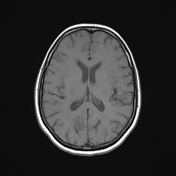

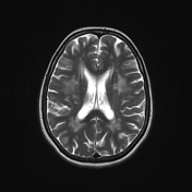


MRI demonstrates extensive hyperintense areas involving supratentorial white matter, subcortical, periventricular and, especially and symmetrically, bitemporal on T2 and FLAIR weighted imaging.
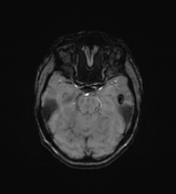
SWI shows a small signal loss area at the left temporal lobe, probably secondary to microhemorrhage.
There is no significant associated brain atrophy.
Case Discussion
MRI is an important diagnostic tool and can predict prognosis of CADASIL, since was established a direct relation between the load of lesions at baseline and the speed of lesions progression at follow-up. Patients with a high MR imaging lesion load are at risk for faster progression of the disease, so they may require more frequent clinical monitoring and MR imaging follow-up.


 Unable to process the form. Check for errors and try again.