
Presentation
Left proptosis and reduced vision.
Patient Data
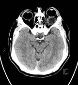

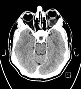

Gross left-sided proptosis with a low-density intracoronal mass lesion within the left orbit. The optic nerve is stretched around the medial aspect of the lesion which is predominantly of low density measuring approximately 10 HU in density. An enhancing component is seen in the inferior and medial margins. The lesion appears largely fluid-filled, though the enhancing component is concerning for tumor.
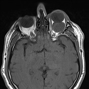
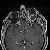
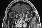
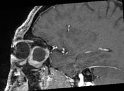
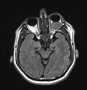

Mixed solid and cystic intraconal mass in the left orbit with associated orbital proptosis. The differential would include both benign and malignant lesions, however, there is the suggestion of bony remodeling reflecting a slow growing/longstanding process, which is more supportive of the former. Other differentials would include nerve sheath tumor. Metastasis would be less likely given the interval stability.
Case Discussion
This patient presented with count-fingers vision in the left eye, after noticing reduced vision for over 1 year. The patient underwent left orbitotomy where the well-circumscribed nodule with a smooth and glistening lining was successfully removed. Histopathology confirmed a benign schwannoma.


 Unable to process the form. Check for errors and try again.