
Presentation
Pelvic pain.
Patient Data
Age: 40 years
Gender: Female
From the case:
Pelvic hydatid cyst


Download
Info
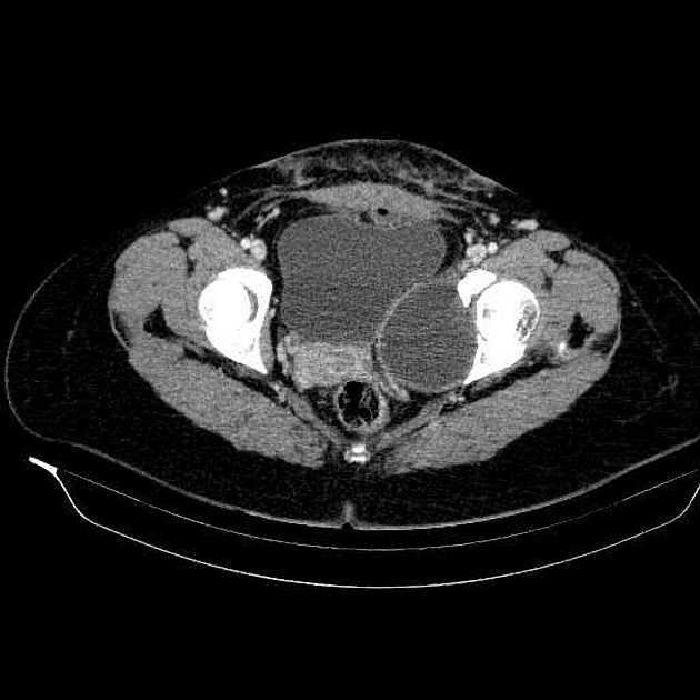
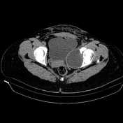
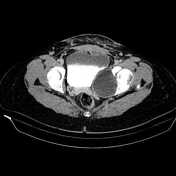
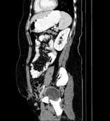
An 83×78×73mm thick-walled cystic lesion is noted at left lower pelvis which extends from lower aspect of left ovary until level of obturator muscles. No soft tissue component is present within the cyst. The lesion erodes medial aspect of left acetabulum but there is no CT-detectable intra-articular extension. Fat plane between the lesion and adjacent pelvic structures is intact. A little amount free fluid is noted at posterior cul-de-sac.
Case Discussion
Large pelvic cystic lesion with an adjacent acetabular defect. Hydatid cyst is the main diagnosis, subsequently proven.


 Unable to process the form. Check for errors and try again.