
From the case:
Pterygopalatine fossa - 3D printed model
Download
Info
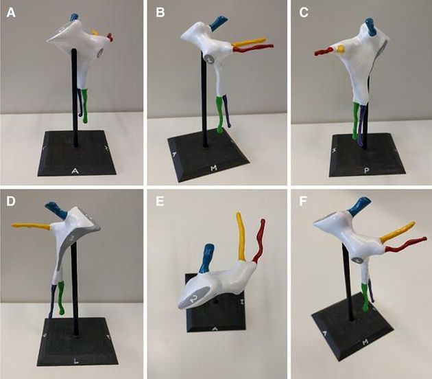
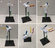
A anterior view. B medial view. C posterior view. D lateral view. E superior view F anterosuperomedial view.
- blue – foramen rotundum
- yellow – pterygoid canal
- red – palatovaginal canal
- green – greater palatine canal
- pink - lesser palatine canal
- sphenopalatine foramen
- inferior orbital fissure
- pterygomaxillary fissure
Case Discussion
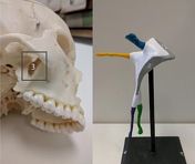
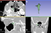
With the advent of 3D printing, a new way of looking at anatomy has been brought into light within medicine. The pterygopalatine fossa (PPF) is a complex anatomical region where the contents and their relationships are usually hard to comprehend and visualize. For this reason, we developed this negative space 3D model of the right PPF, mounted on the bespoke stand. This is the only model that is able to physically conceptualize this interesting space!
Permission was granted from the authors and the respective journal of which this study was published.


 Unable to process the form. Check for errors and try again.